


Orthodontic X Rays – What They Show and Why They Matter
Posted on September 23, 2026
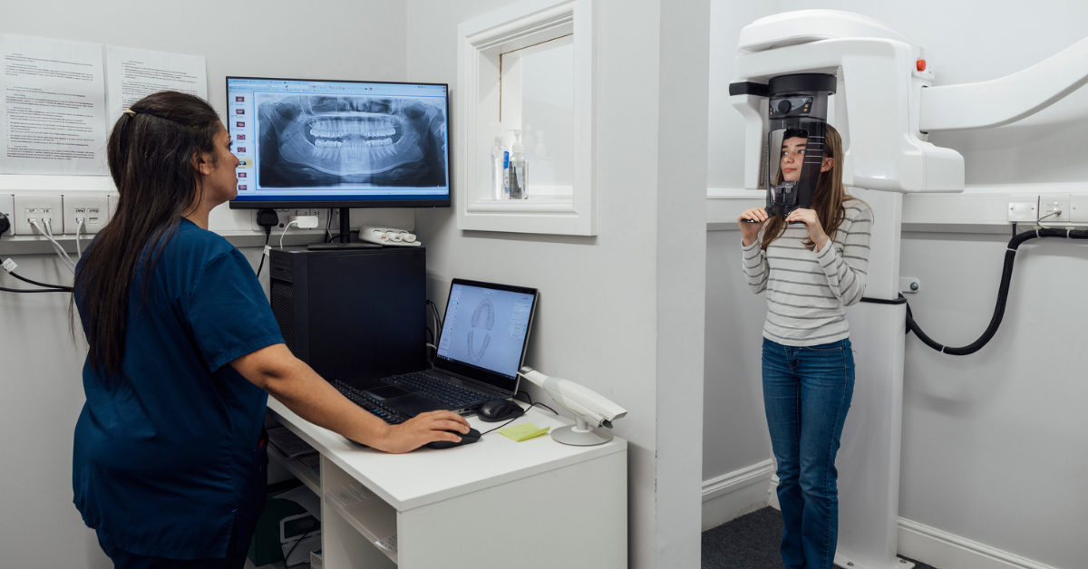
If your orthodontist can already see your teeth, you may wonder why X rays are needed before braces or Invisalign. The answer is that an orthodontic exam only shows part of the picture. Important structures such as tooth roots, developing teeth, impacted teeth, and parts of the jaw are located below the gums and cannot always be fully evaluated by looking inside the mouth.
Orthodontic X rays can provide valuable information that helps an orthodontist understand how the teeth and jaws are developing and how treatment should be planned.
At TruSmile Orthodontics in Long Beach, California, imaging is considered alongside a full orthodontic evaluation. Patients from across Long Beach and Los Angeles County can receive personalized care based on their individual needs, dental development, bite, and treatment goals.
What Are Orthodontic X Rays?
Orthodontic X rays are images that allow an orthodontist to see structures that are not visible during a regular dental examination.
The images may show the roots of the teeth, the jawbone, permanent teeth that have not erupted, and the relationship between the upper and lower jaws. They can also help identify teeth that are missing, extra, impacted, or developing in unexpected positions.
The word radiograph is often used to describe the actual image produced by X ray technology.
For orthodontic patients, these images are only one part of the diagnostic process. Your orthodontist will also consider the appearance of the teeth, the way the bite fits together, digital scans, photographs, dental history, and other clinical information.
Why Are X Rays Important Before Braces or Invisalign?
Orthodontic treatment involves moving teeth through the supporting bone. Before beginning treatment, an orthodontist needs to understand what is happening above and below the gumline.
X rays may help reveal the exact position of tooth roots and show whether permanent teeth are erupting normally. They can also identify impacted teeth that are blocked from coming into the mouth.
For children and teens, imaging may provide information about teeth that are still developing. For adults, it may help the orthodontist understand existing root positions, bone relationships, and previous dental treatment.
The need for X rays should be determined individually. Not every patient requires the same images, and not every patient needs imaging at the same time.
What Can an Orthodontist See on an X Ray?
Orthodontic X rays can provide information that supports diagnosis and treatment planning.
Tooth Roots
The crown is the part of the tooth you see when you smile, but each tooth also has a root beneath the gumline. Knowing the position and shape of the roots can help an orthodontist plan tooth movement.
Developing Permanent Teeth
Children and teens may have permanent teeth that have not erupted yet. X rays can show where these teeth are developing and whether they appear to be following an expected eruption path.
Impacted Teeth
An impacted tooth is a tooth that cannot erupt properly into the mouth. Canine teeth are one example of teeth that may become impacted.
Finding an impacted tooth can help the orthodontist determine whether additional treatment may be needed.
Missing or Extra Teeth
Some people are born without certain permanent teeth. Others develop extra teeth. These conditions may not always be obvious during a visual examination.
Imaging can help identify these situations and support a treatment plan that considers spacing and bite alignment.
Jaw and Bone Relationships
Certain images allow orthodontists to evaluate the relationship between the teeth and jaws. This may be helpful when planning treatment for an overbite, underbite, crossbite, or another alignment concern.
Types of X Rays Used in Orthodontics
Different types of images serve different purposes. An orthodontist will determine which images, if any, are appropriate based on the patient and the information needed.
Panoramic X Ray
A panoramic X ray provides a broad view of the teeth and jaws in a single image.
It may show erupted teeth, developing teeth, tooth roots, impacted teeth, and general jaw structures. This type of image can be especially helpful when an orthodontist needs to understand how multiple teeth are positioned at the same time.
Cephalometric X Ray
A cephalometric X ray usually provides a side view of the head.
Orthodontists may use this type of image to study the relationship between the teeth, jaws, and facial structures. It can be useful when evaluating bite relationships and planning orthodontic treatment.
Periapical X Ray
A periapical X ray focuses on a smaller area.
It shows the entire tooth, including the root and surrounding bone. An orthodontist may request this type of image when more detailed information about a specific tooth is needed.
Bitewing X Rays
Bitewing X rays are often taken during general dental visits. They are commonly used to evaluate the areas between teeth and look for signs of decay.
An orthodontist may review existing bitewing images when they provide useful information for treatment planning.
CBCT Imaging
Cone beam computed tomography, commonly called CBCT, creates a three dimensional image.
A CBCT scan may be useful in selected cases involving impacted teeth, complicated root relationships, jaw concerns, or other situations where additional information is needed.
CBCT imaging is not necessary for every orthodontic patient. It is generally reserved for cases where the added detail may affect diagnosis or treatment planning.
Are Orthodontic X Rays Safe?
Questions about radiation are common among patients and parents.
Dental X rays use ionizing radiation. Because radiation exposure should be limited whenever possible, imaging should be ordered when it provides meaningful clinical information.
Modern digital imaging technology is designed to produce useful images while managing exposure. The type of image, the patient’s size, and the information needed all influence how imaging is selected.
It is also important to understand that orthodontic X rays are not taken simply because a certain amount of time has passed. The decision should be based on the patient’s individual needs and treatment progress.
Patients at TruSmile Orthodontics can ask questions about why an image is recommended and how it will be used in their treatment plan.
Are Dental X Rays Safe for Children?
Parents often have additional questions when X rays are recommended for a child.
Children may need imaging to evaluate permanent teeth that are still developing, eruption patterns, crowding, impacted teeth, or bite concerns.
Because children are still growing, the orthodontist considers age, size, dental development, and the specific information needed when selecting an imaging method.
An early orthodontic evaluation does not mean a child automatically needs X rays or braces. In some cases, the orthodontist may recommend monitoring development before beginning treatment.
How Often Do You Need X Rays During Orthodontic Treatment?
There is no universal X ray schedule that applies to every orthodontic patient.
Some patients may need imaging before treatment begins and again later to evaluate certain changes. Other patients may require fewer images.
The frequency can depend on factors such as age, tooth eruption, treatment complexity, root position, and specific clinical concerns.
An orthodontist may also review existing X rays from a dentist before deciding whether additional images are needed.
The goal is to obtain the information needed for safe and effective treatment planning without taking unnecessary images.
Can TruSmile Use X Rays From My Dentist?
In some cases, yes.
If your general dentist recently took dental X rays, those images may provide useful information for your orthodontic evaluation. It can be helpful to share existing images with the orthodontist when possible.
The orthodontist will review the images and determine whether they provide the views and details needed for orthodontic planning.
Additional imaging may still be recommended if the existing X rays do not show the information needed for treatment.
This approach helps avoid repeating images when suitable recent records are already available.
What Happens When You Get an Orthodontic X Ray?
The process depends on the type of X ray being taken.
For a panoramic X ray, the patient will usually stand or sit while the imaging machine moves around the head. You may be asked to bite gently on a positioning guide and remain still while the image is captured.
You may also be asked to remove earrings, necklaces, glasses, or other metal items that could interfere with the image.
The process is generally quick and does not involve injections or other invasive procedures.
Once the image is available, the orthodontist can review it along with the rest of your diagnostic records.
How X Rays Help Create a Personalized Treatment Plan
X rays do not determine treatment by themselves.
They are considered alongside a complete orthodontic examination, photographs, digital scans, bite evaluation, dental history, and the patient’s goals.
For example, two patients may both have crowded front teeth but require different treatment approaches because their roots, jaw relationships, or developing teeth are different.
Imaging can help the orthodontist understand these differences before treatment begins.
At TruSmile Orthodontics, treatment planning is customized to the individual patient rather than based only on appearance.
X Rays for Braces vs Invisalign
Patients sometimes wonder whether X ray needs are different for braces and Invisalign.
The type of appliance does not determine the full diagnostic process. Whether you are considering braces or clear aligners, your orthodontist still needs to understand the position of the teeth, roots, and jaws.
Braces and Invisalign move teeth in different ways, but both require thoughtful planning.
The orthodontist will decide which images are appropriate based on your anatomy and treatment needs rather than simply on which appliance you prefer.
What to Expect at Your TruSmile Orthodontic Consultation
Your first orthodontic visit is an opportunity to understand your smile and learn about possible treatment options.
The orthodontic team will typically begin by reviewing your dental and health history. Your teeth and bite will then be examined to identify crowding, spacing, bite problems, or other concerns.
If diagnostic records are needed, the orthodontist may recommend photographs, digital scans, or X rays.
Once enough information has been gathered, the orthodontist can explain your findings and discuss possible treatment choices such as braces, clear aligners, or other orthodontic appliances.
You can also ask questions about treatment time, dental insurance, financing, and next steps.
Making Orthodontic Care More Affordable
Cost is an important consideration for many families in Long Beach and throughout Los Angeles County.
TruSmile Orthodontics works to make orthodontic treatment more accessible through several payment options.
The practice accepts most PPO plans, HMOs, and Denti Cal. Customizable financing is available, along with zero percent financing options for qualifying patients and affordable monthly payment plans. CareCredit is also accepted.
Once your treatment needs are understood, the TruSmile team can review costs and financial options with you.
Why Choose TruSmile Orthodontics in Long Beach?
Orthodontic treatment begins with understanding what is happening with your teeth and bite.
TruSmile Orthodontics provides personalized care for children, teens, and adults in Long Beach, California. Modern diagnostic tools, including orthodontic imaging when appropriate, help support careful treatment planning.
Patients can explore braces, clear aligners, and other treatment options in a welcoming environment where questions are encouraged.
The practice also offers flexible financial arrangements and serves families throughout Long Beach and Los Angeles County.
Schedule an Orthodontic Consultation in Long Beach
Orthodontic X rays can provide information about tooth roots, developing teeth, impacted teeth, jaw relationships, and other structures that cannot always be fully evaluated during a visual examination.
The type and timing of imaging should be based on the individual patient rather than a standard schedule.
If you are considering braces, Invisalign, or another orthodontic treatment, schedule a consultation with TruSmile Orthodontics in Long Beach and Culver City, California. Our team can evaluate your smile, explain whether orthodontic X rays are appropriate, and create a personalized treatment plan based on your needs.
TruSmile Orthodontics proudly serves children, teens, adults, and families from Long Beach, Culver City and communities throughout Los Angeles County.
